Best Imaging For Orbital Floor And Maxillary Fracture

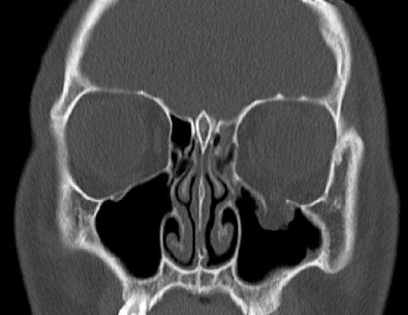
Blowout Fracture Of The Orbital Floor Ct Of The Midface Frontal View Bone Window Orbital O Floor Fr In 2020 Rectus Muscle This Or That Questions Maxillary Sinus
Wrenching Up The Socket
Orbital Blowout Fracture Radiology At St Vincent S University Hospital

Imaging The Face Radiology Key

Delayed Onset Inferior Rectus Muscle Hematoma After Orbital Floor Fracture Jama Ophthalmol 2013 131 11 1492 Doi 10 1001 Jamao Rectus Muscle Fracture Muscle

Orbital Floor Fracture Radiology Case Radiopaedia Org

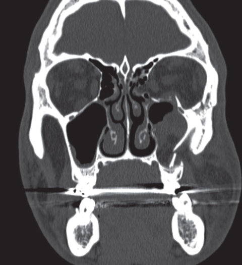
The floor is usually the path of least resistance and fractures downward into the maxillary sinus.
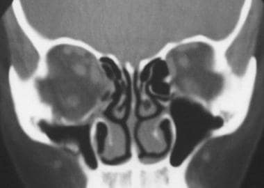
Best imaging for orbital floor and maxillary fracture. This patient had a significant vertical ocular motility disturbance. The usual mechanism is a blow to the eye with the forces being transmitted by the soft tissues of the orbit downward to the thin floor of the orbit. Orbital floor fracture also known as blowout fracture of the orbit eye socket. Mid face le fort fractures.
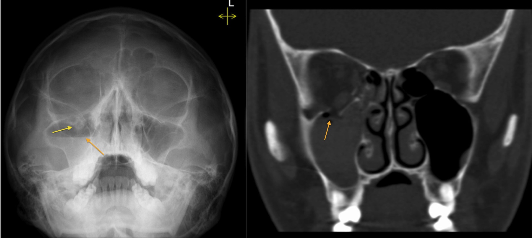
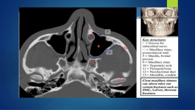
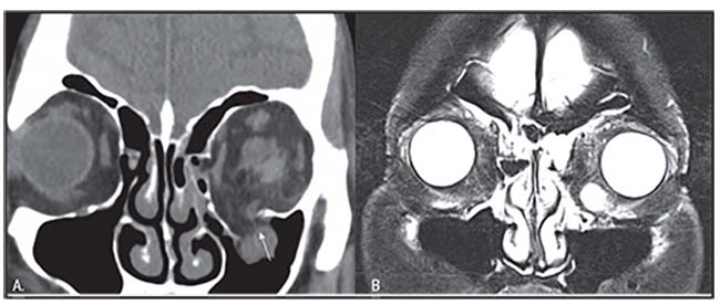
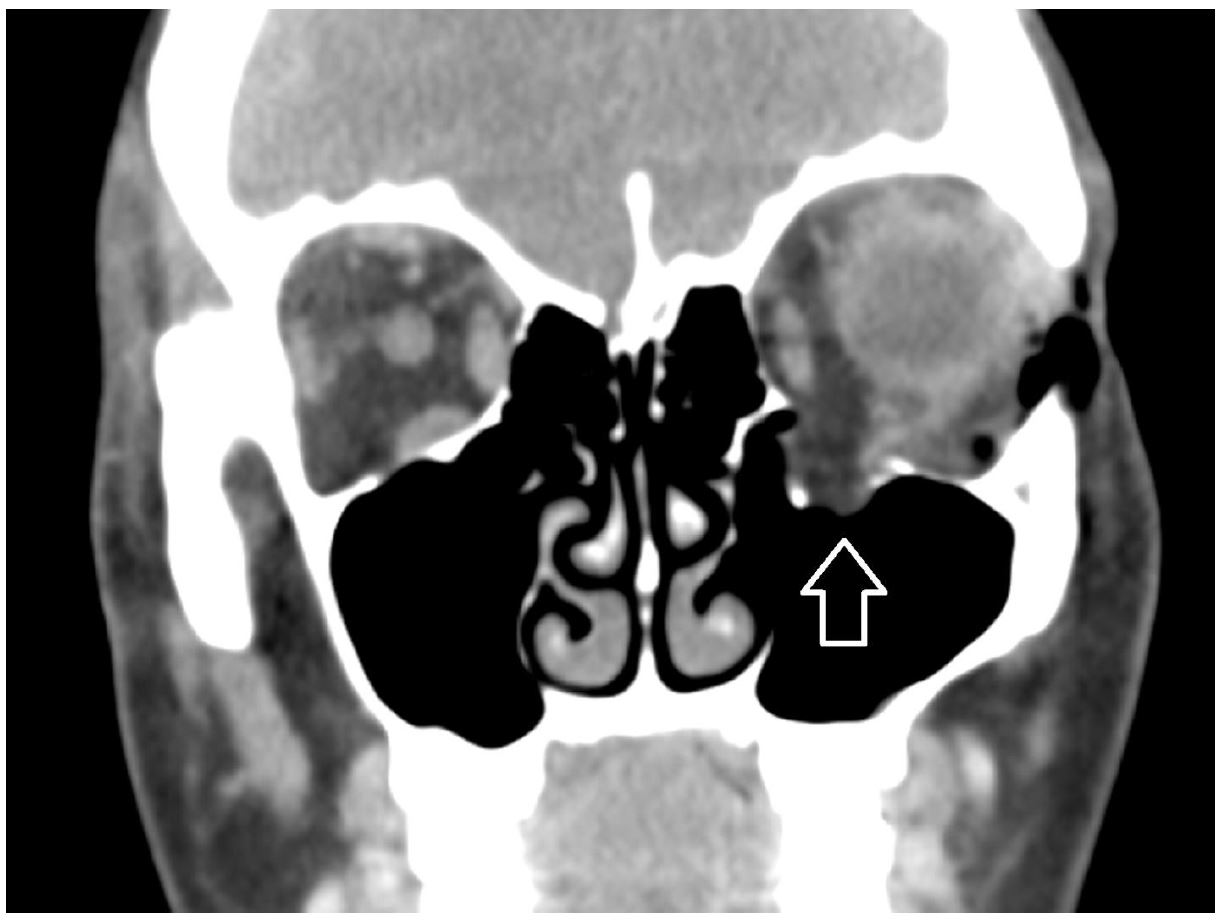
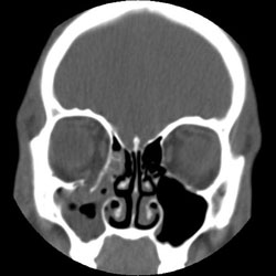
The orbit is formed by 7 bones. Blunt force trauma tends to cause fractures along three lines of weakness in the mid face. Note the relatively small amount of herniated tissue and the air fluid level in the maxillary sinus. A retrospective study by bartoli et al of 301 orbital floor fractures found the most common symptom to be hypesthesia extending through the region of the maxillary nerve 32 9 of patients.
Fractures of the orbit may be seen in different scenarios of direct and indirect trauma to the globe orbital facial or cranial bones. One characteristic of all types of le fort fractures is the fracture of the. Orbital floor fractures may result when a blunt object which is of equal or greater diameter than the orbital aperture strikes the eye or on the cheek 1. Another common fracture is the orbital floor fracture or blowout fracture.
Getting hit with a baseball or a fist often causes a orbital blowout fracture. Direct orbital floor fracture. Inferior floor medial wall lamina papyracea superior roof lateral wall. Inferior blowout fractures are the most common.
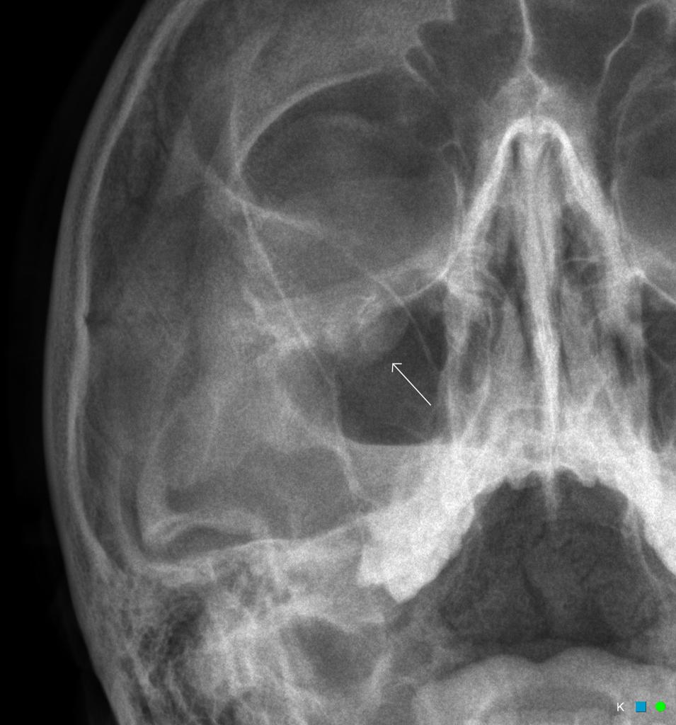
Maxillary bones upper jaw. This is a rim fracture that extends into the lower socket. Orbital floor fracture with significant soft tissue entrapment a so called trapdoor fracture. Waters view best displays inferior orbital rims nasoethmoidal bones and maxillary sinuses.
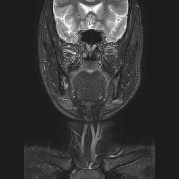
A study by huang et al indicated that in patients with head trauma lack of maxillary hemosinus on conventional head ct scanning predicts the absence of orbital floor fracture the negative. If the patient is upright when the film is taken an air fluid level can often be seen in the maxillary sinus which may indicate fracture of the maxillary sinus orbital floor. Orbital fat prolapses into the maxillary sinus and may be joined by prolapse of the inferior rectus muscle. Zygomatic sphenoid maxillary frontal lacrimal palatine and ethmoid.
Blowout fractures can occur through one or more of the orbital walls.

Learningradiology Facial Bones Rectus Muscle Maxillary Sinus

Maxillary Sinus Abnormal Maxillary Sinus Sinusitis Radiology Imaging
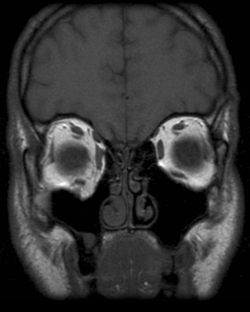
Coronal T1 Mri Scan Of A Right Orbital Floor Fracture A The Black Download Scientific Diagram
Imaging In Facial Trauma

Pin De Patrick Caulfield En Dark En 2020

Pin On Ct Scans

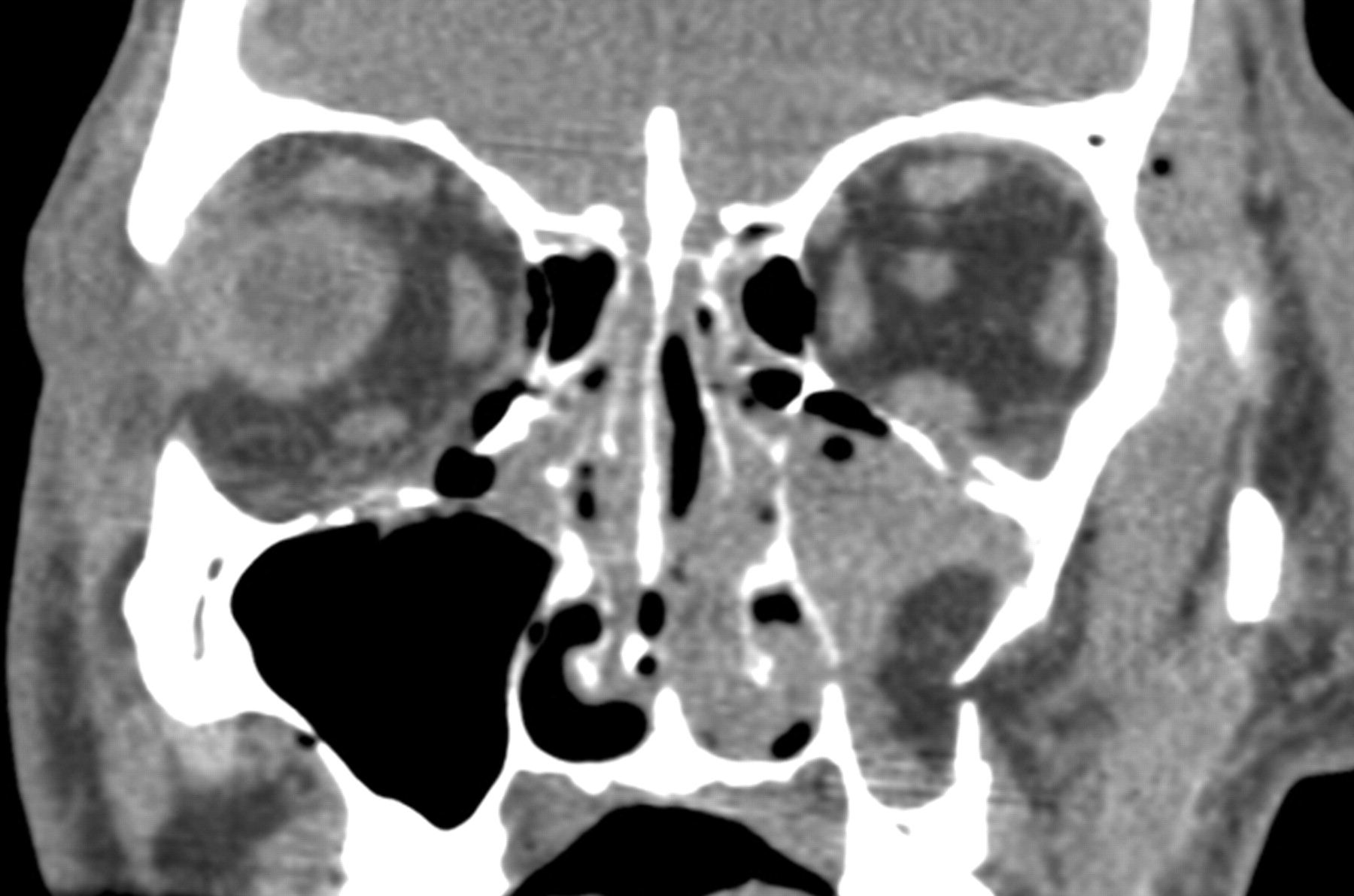
Coronal Ct Imaging Of A Pediatric Patient With A Left Orbital Floor Download Scientific Diagram
Orbital Blow Out Fracture Mri Radiology Case Radiopaedia Org
December 2018 Wills Eye Resident Case Series Diagnosis Discussion
Blow Out Fracture Right Orbital Floor Radiology Case Radiopaedia Org

Pin On Ct Scans
The Measurement Of Orbital Blowout Fractures Cannot Be Made With Geometric Estimations

Ct Imaging Of The Face Chapter 31 Clinical Emergency Radiology

Black Eyebrow Sign Black Eyebrow Sign Is The Description Given On Plain Facial Radiographs To Intra Orbital Air Air Rises Black Eyebrows Radiology Air Leaks

Trauma Radiology Key

Fibrous Dysplasia Of Skull Base Radiology Bone Diseases Head And Neck

Full Text Management Of Orbital Fractures Challenges And Solutions Opth

Facial And Mandibular Fractures Uw Radiology Fractures Radiology Complex Fracture
Https Encrypted Tbn0 Gstatic Com Images Q Tbn 3aand9gcssxho1wdwxhgszomrieyincvjfsjyupzte5vsui1liqnjcljxl Usqp Cau

Orbital Floor Blow Out Fracture Types Of Fractures Maxillary Sinus Nursing Information

Orbital Pseudotumour Radiology Reference Article Radiopaedia Org Radiology Head And Neck Cat Scan

Complex Orbital Fracture Repair Using Rigid Fixation Of The Internal Orbital Skeleton World Renowned Bespoke Cosmetic Plastic Surgeon Boston Dr Michael Yaremchuk

Teardrop Sign Of Orbital Blowout Fracture Radiology Case Radiopaedia Org
Orbital Blowout Fracture From Nose Blowing The Western Journal Of Emergency Medicine

Pin On Mri

3 Head And Neck Radiology Key

Fracture Pattern Of The Zygoma Red Infraorbital Fracture Site Turquois Orbital Floor Fracture Site Blue Fracture Site Of Th Skull Tattoo Pattern Cat Scan

Pin On Ent Rotation

Pin On Kb

Facial Trauma Undergraduate Diagnostic Imaging Fundamentals

6 Orbit And Globe Table 6 3 Table 6 4 Radiology Key

Paranasal Sinus Fractures Radiology Reference Article Radiopaedia Org

Invasive Fungal Sinusitis Radiology Case Radiopaedia Org Radiology Radiology Imaging Sinusitis
Brown Emergency Medicine
Trauma Service Maxillofacial Injury
Orbital Blowout Fracture Radiology Reference Article Radiopaedia Org

Pin By Dr Abuaiad On Brain Head And Neck Head And Neck Floor Show Tongue Piercing
Herniation Of The Buccal Fat Pad Into The Maxillary Antrum Ct Findings In Three Cases American Journal Of Neuroradiology
Orbital Floor Fractures Blowout Workup Laboratory Studies Imaging Studies Other Tests

Sphenoethmoidal Air Cell Radiology Reference Article Radiopaedia Org Radiology Cell Internal Carotid Artery

Orbit Blunt Force Fractures And Penetrating Injuries Radiology Key

Aneurysmal Bone Cyst Radiology Case Radiopaedia Org Hand Therapy Cysts Radiology
